Back Muscles Anatomy Labeled : Muscles Of The Back - Human muscle system, the muscles of the human body that work the skeletal system, that are under voluntary control, and that are concerned with the following sections provide a basic framework for the understanding of gross human muscular anatomy, with descriptions of the large muscle groups.
Back Muscles Anatomy Labeled : Muscles Of The Back - Human muscle system, the muscles of the human body that work the skeletal system, that are under voluntary control, and that are concerned with the following sections provide a basic framework for the understanding of gross human muscular anatomy, with descriptions of the large muscle groups.. Understanding the anatomy and function of your back muscles can help you determine if (and when) you may need professional medical care if you are. Anatomy of the muscular system chapter 10 281. Learn anatomy faster and remember everything you learn. Study flashcards on anatomy back muscles at cram.com. The muscular system is responsible for movement in collaboration with the nervous system to form impulses for motion.
Muscles of the back can be divided into superficial, intermediate, and deep group. Learn about these muscles, their locations there are several individual muscles within the back anatomy, and it's important to take a quick look at all of them to see how you can target them. Related posts of muscles labeled front and back. This is my video about the muscles of the back. Muscles, connected to bones or internal organs and blood vessels, are in charge for movement.
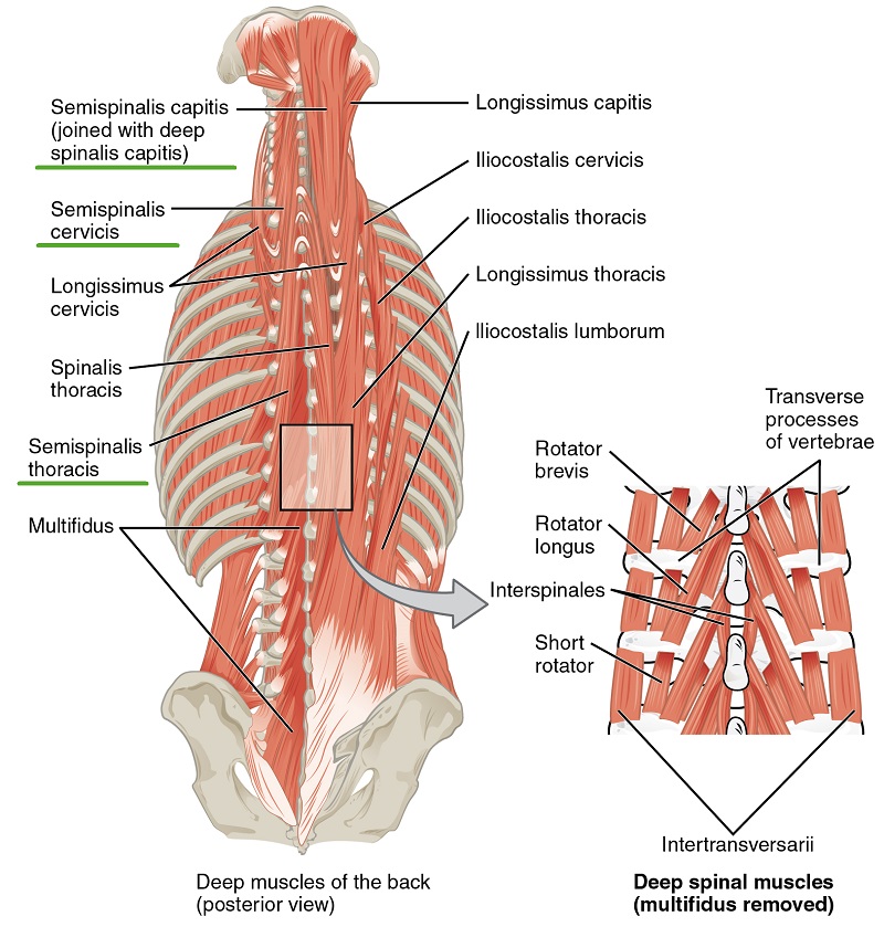
Intrinsic Back Muscles Anatomy Of The Torso Medical Library from d3uigcfkiiww0g.cloudfront.net Muscles of the back can be divided into superficial, intermediate, and deep group. Learn about the superficial, intermediate and deep muscles of the back. Learn about these muscles, their locations there are several individual muscles within the back anatomy, and it's important to take a quick look at all of them to see how you can target them. Their main function is contractibility. The extrinsic muscles include the trapezius, latissimus dorsi, rhomboid major and minor, levator scapulae and the serratus posterior superior and. They can be affected by various conditions. There are around 650 skeletal muscles within the typical human body. Learn about anatomy muscle labeling with free interactive flashcards.
3d interactive modules and video tutorials on the anatomy of the back muscles.
Labels are a means of identifying a product or container through a piece of fabric, paper, metal or plastic film onto which information about them is printed. The extrinsic muscles include the trapezius, latissimus dorsi, rhomboid major and minor, levator scapulae and the serratus posterior superior and. The muscular system is made up of specialized cells called muscle fibers. Memorize all the muscle facts with the help of muscle cheat sheets. Tutorials on the anatomy and actions of the back muscles, using interactive animations, diagrams, and illustrations. Click on the labels below to find out more about your muscles. The extrinsic back muscles are also referred to as secondary back muscles. 3d interactive modules and video tutorials on the anatomy of the back muscles. Muscles also contribute to internal functions of the human body which include motion in the intestines and circulatory system. The muscular system is responsible for movement in collaboration with the nervous system to form impulses for motion. Topographically, the muscles in this group are classed along with the lateral torso wall and upper extremity, which is due to their location as well as their genetic development based on their embryological origin. There are around 650 skeletal muscles within the typical human body. This article looks at the anatomy of the back, including bones, muscles, and nerves.
Muscles of the back can be divided into superficial, intermediate, and deep group. Intermediate back muscles and c. Labels are a means of identifying a product or container through a piece of fabric, paper, metal or plastic film onto which information about them is printed. The manner in which muscles are grouped, the relationship of muscles to joints. The extrinsic back muscles are also referred to as secondary back muscles.
Muscles Of The Neck And Torso Classic Human Anatomy In Motion The Artist S Guide To The Dynamics Of Figure Drawing from doctorlib.info Exercise of this organ system is critical to prevent. By jholmesrn419 , may 2007. Muscle basics and cellular components, naming of the muscles, and cat. This quiz requires labeling, so it will test your knowledge on how to identify these muscles (latissimus dorsi, trapezius, deltoid, biceps brachii. Learn about these muscles, their locations there are several individual muscles within the back anatomy, and it's important to take a quick look at all of them to see how you can target them. Muscles of the back can be divided into superficial, intermediate, and deep group. Labels are a means of identifying a product or container through a piece of fabric, paper, metal or plastic film onto which information about them is printed. This is my video about the muscles of the back.
Muscle basics and cellular components, naming of the muscles, and cat. The extrinsic muscles that are associated with upper extremity and shoulder movement, and the intrinsic injuries of the intrinsic back muscles often occur while using improper lifting technique. This article looks at the anatomy of the back, including bones, muscles, and nerves. Memorize all the muscle facts with the help of muscle cheat sheets. It also covers some common conditions and injuries that can affect the. The intrinsic back muscles, which are also called true back muscles. Neck muscle anatomy mri 12 photos of the neck muscle anatomy mri neck muscle anatomy images, neck muscle anatomy pictures, neck muscle anatomy posterior, neck muscle anatomy ultrasound, neck muscles anatomy radiology. The superficial back muscles are the muscles found just under the skin. Muscles also contribute to internal functions of the human body which include motion in the intestines and circulatory system. This site was designed for students of anatomy and physiology. Back muscles are divided into two specific groups: This chapter is divided into three main sections: The back muscles can be three types.
Learn about the superficial, intermediate and deep muscles of the back. When you are taking anatomy and physiology you will be required to identify major muscles in the human body. Learn about anatomy muscle labeling with free interactive flashcards. The muscles of the back are separated into extrinsic and intrinsic components, which are based on their function in movement and embryological origin. Muscles of the back can be divided into superficial, intermediate, and deep group.
Beschriftete Anatomiechart Von Mannlichen Trizepten Und Ruckenmuskeln Auf Weissem Hintergrund Stockfoto Und Mehr Bilder Von Anatomie Istock from media.istockphoto.com Anatomical diagram showing a back view of muscles in the human body. Topographically, the muscles in this group are classed along with the lateral torso wall and upper extremity, which is due to their location as well as their genetic development based on their embryological origin. Muscles also contribute to internal functions of the human body which include motion in the intestines and circulatory system. This site was designed for students of anatomy and physiology. Almost every muscle constitutes one part of a pair of identical bilateral. This is my video about the muscles of the back. Their main function is contractibility. This article looks at the anatomy of the back, including bones, muscles, and nerves.
Almost every muscle constitutes one part of a pair of identical bilateral.
Rotator cuff muscles parts pinterest bones body diagram elegant labeled skeleton back view male dog muscular system science pinterest of biceps femoris tendons 751 1300—1335 sternothyroid muscle anatomy function & diagram les 8 meilleures images du tableau jack sur pinterest endocrine system. Their main function is contractibility. This site was designed for students of anatomy and physiology. The muscles of the back that work together to support the spine, help keep the body upright and allow twist and bend in many directions. Anatomy of the muscular system chapter 10 281. Exercise of this organ system is critical to prevent. Topographically, the muscles in this group are classed along with the lateral torso wall and upper extremity, which is due to their location as well as their genetic development based on their embryological origin. The extrinsic muscles that are associated with upper extremity and shoulder movement, and the intrinsic injuries of the intrinsic back muscles often occur while using improper lifting technique. Click on the labels below to find out more about your muscles. The extrinsic muscles include the trapezius, latissimus dorsi, rhomboid major and minor, levator scapulae and the serratus posterior superior and. Microscopic anatomy of skeletal muscle. They can be affected by various conditions. Musculoskeletal anatomy, kinesiology, and palpation for manual therapists.
Learn about the superficial, intermediate and deep muscles of the back back muscles anatomy. Clearly pictured and labeled muscles of the back and neck.


Posting Komentar
0 Komentar